Swati — Medical Radiology & Imaging Technology
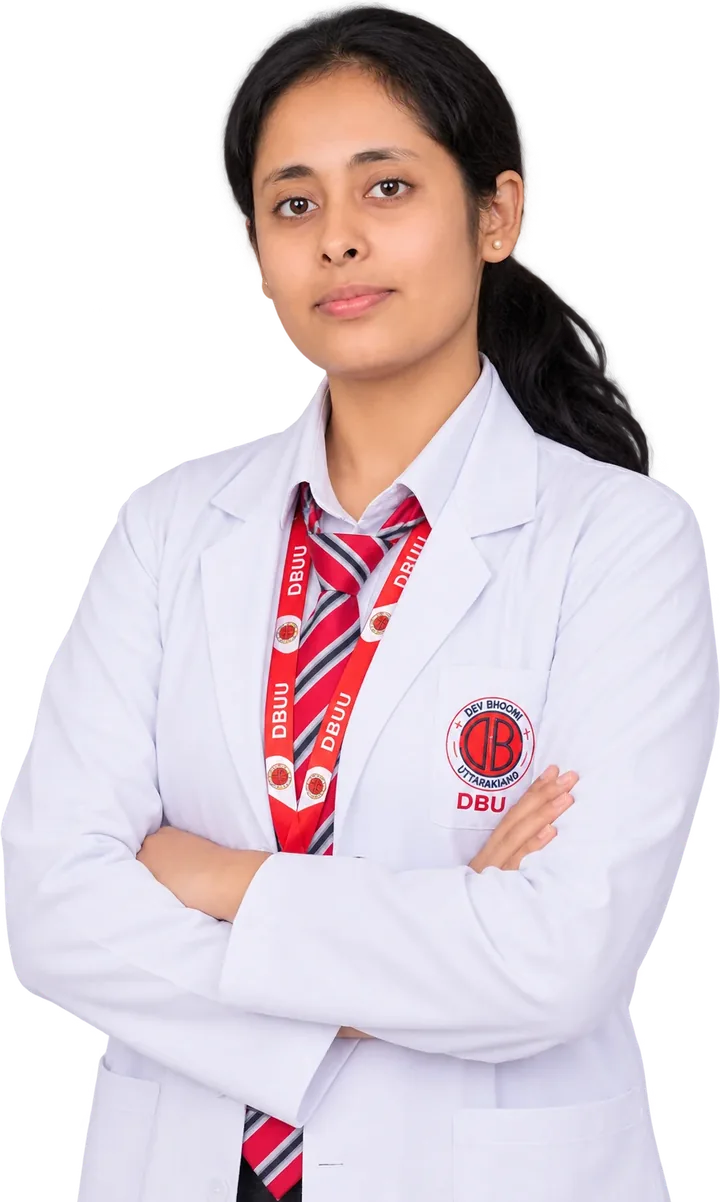
I'm Swati, a BMRIT student at Dev Bhoomi Uttarakhand University, Dehradun — training in radiography, CT and MRI with a focus on accurate positioning, radiation safety and calm, patient-first care.
- Programme
- BMRIT · Year 2, Sem 3
- University
- Dev Bhoomi Uttarakhand
- Focus
- Radiography · CT · MRI
- Based in
- Dehradun, India
- Digital Radiography
- Computed Tomography
- MRI Fundamentals
- Radiation Protection
- Patient Positioning
- Sectional Anatomy
- Imaging Physics
- Patient-first Care
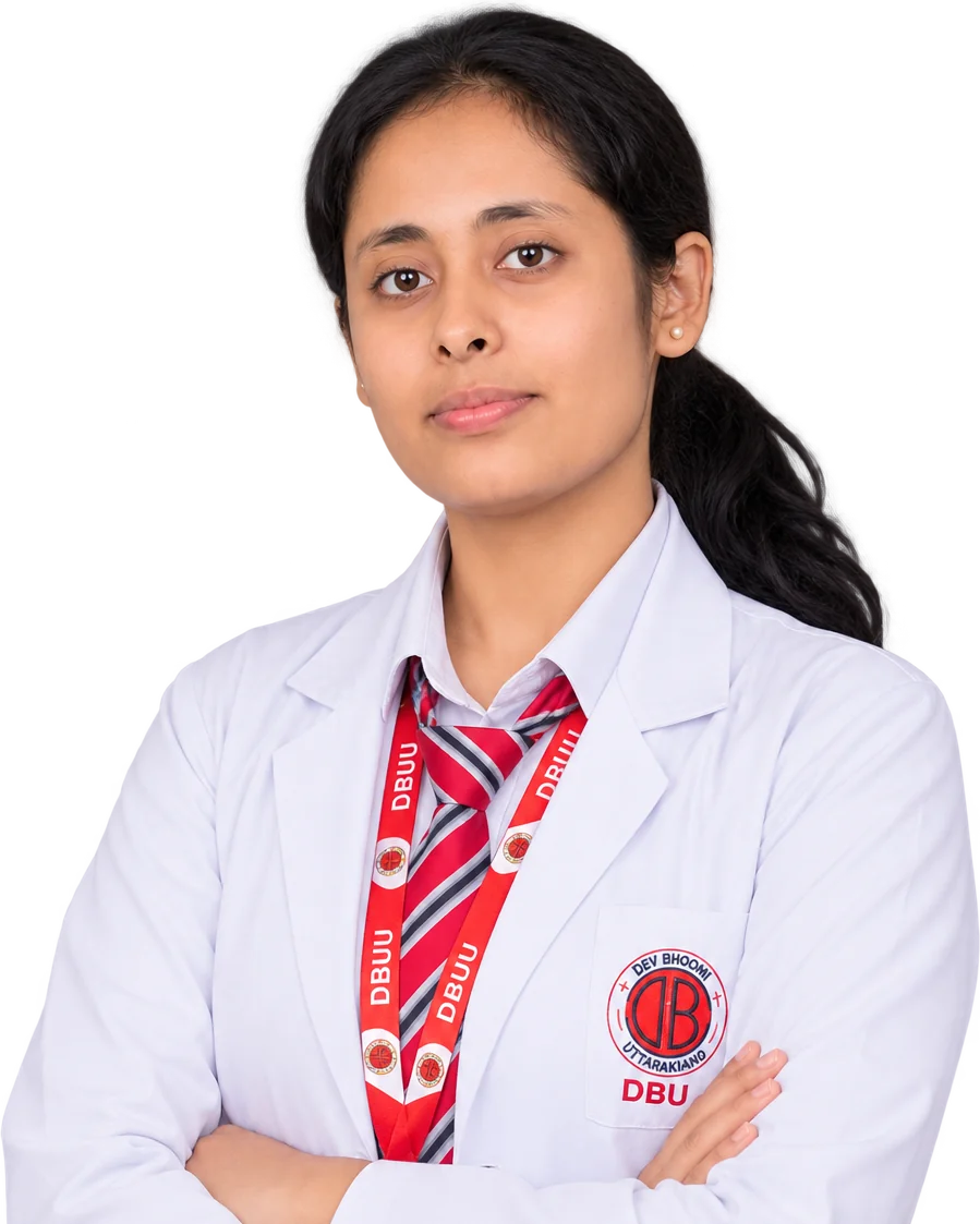
I believe a great image starts long before the exposure — with a patient who feels safe, a position that is precise, and a dose that is only ever as much as it needs to be.
I'm a Bachelor of Medical Radiology & Imaging Technology student at Dev Bhoomi Uttarakhand University. Alongside coursework in anatomy, radiation physics and radiographic technique, I've trained in positioning labs, observed digital radiography and CT in a working radiology department, and attended workshops on CT/MRI fundamentals and radiation protection.
- Programme
- Bachelor of Medical Radiology & Imaging Technology
- Current
- Year 2 · Semester 3
- University
- Dev Bhoomi Uttarakhand University, Dehradun
- Languages
- English (professional) · Hindi (native)
Year of BMRIT
Core subjects in coursework
Imaging modalities studied
From a science-stream schooling in Bihar to a specialised degree in medical radiology and imaging technology.
- 2025 — Present
Bachelor of Medical Radiology & Imaging Technology
Dev Bhoomi Uttarakhand University · Dehradun, Uttarakhand
Building foundations in radiographic technique, radiation physics, sectional anatomy and patient care.
- BMRIT
- Year 2 · Sem 3
- SGPA 8.25
- 2021 · Class XII
Senior Secondary — Medical (PCB)
G.D.S. Academy · Raxaul, East Champaran, Bihar
- CBSE
- 66%
- School code 65196
- 2019 · Class X
Secondary School
Chandrasheel Vidyapeeth · Kanti, Muzaffarpur, Bihar
- CBSE
- 63.8%
- School code 50046
Structured practicals, a working radiology department and focused workshops — building the habits that matter on the floor.
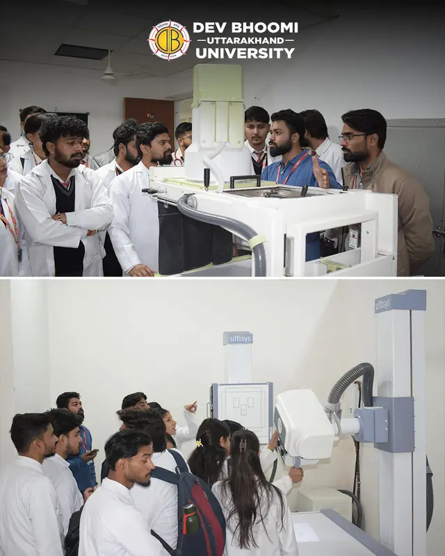
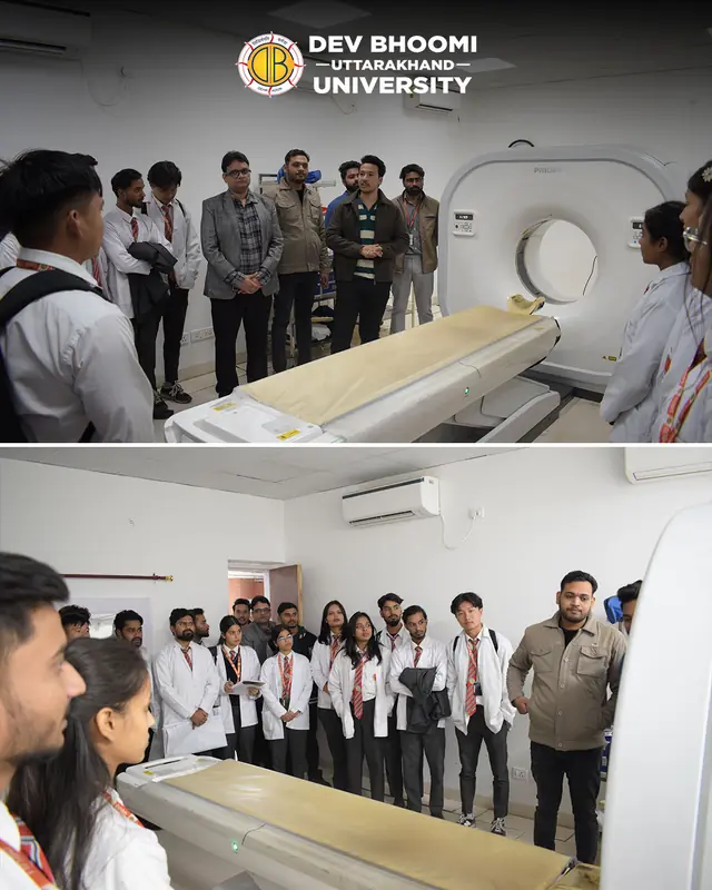
Radiology Department — Educational Visit
Dev Bhoomi Medical College of Paramedical Sciences
Observed a digital radiography suite and a Philips CT scanner in a working radiology department — patient preparation, positioning, console workflow and radiation-safety practice, guided by radiology faculty.
- Digital radiography
- CT
- Clinical workflow
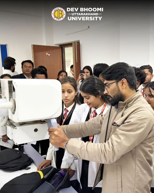
Radiographic Positioning & Imaging Lab
Dev Bhoomi Uttarakhand University
Practised standard X-ray projections, image acquisition and equipment handling under faculty supervision, applying ALARA radiation-protection principles.
- Positioning
- Exposure factors
- ALARA
Imaging & Radiation-Safety Workshops
Departmental workshops
Practical sessions on CT and MRI fundamentals, patient preparation and radiation protection for patients and staff.
- CT
- MRI
- Radiation safety
Seminars & case studies
- Seminar
Advances in Diagnostic Imaging
Presented emerging imaging technologies and their role in earlier, more accurate diagnosis.
- Case study
Radiographic Anatomy
Correlated cross-sectional anatomy with the common radiographic projections used in practice.
The core competencies I'm developing for safe, diagnostic-quality imaging.
Radiographic Procedures
Standard projections for the chest, extremities, spine and skull.
Patient Positioning
Accurate, comfortable positioning for diagnostic-quality images.
Medical Imaging Basics
How X-ray, CT and MRI form an image — and what degrades it.
Radiation Safety
ALARA, shielding and time–distance principles; dose awareness.
Anatomy & Physiology
Systemic and sectional anatomy for confident image reading.
Diagnostic Imaging Fundamentals
Image quality, artefacts and the role of imaging in diagnosis.
Professional skills
- Clear patient communication
- Teamwork
- Leadership
- Time management
- Problem solving
- Empathy
Languages
- EnglishProfessional working
- HindiNative
The subjects that shape how I read an image, position a patient and protect them from unnecessary dose.
- 01Human AnatomySystemic & sectional
- 02Human PhysiologyOrgan systems
- 03PathologyDisease on imaging
- 04Radiographic TechniquesProjections & exposure
- 05Radiation PhysicsX-ray production & interaction
- 06Medical EthicsConsent, dignity, confidentiality
- 07Imaging Equipment BasicsDR, CT & MRI hardware
My BMRIT grades so far, and the activities I've taken part in outside the lecture hall.
Academic record · SGPA out of 10
Semester 1
0.00Semester 2
0.00Semester 3
In progress
Activities
Imaging & radiation-safety workshops
Hands-on sessions on CT and MRI fundamentals, patient preparation and radiation protection.
Seminar presentation
Presented on advances in diagnostic imaging and their role in earlier diagnosis.
Anatomy case study
Correlated cross-sectional anatomy with common radiographic projections.
Health-camp volunteering
Supported community health camps and awareness drives on preventive care.
Radiology-department visits, practicals and life on campus at Dev Bhoomi Uttarakhand University.
Open to clinical internships, observerships and trainee opportunities in radiology departments. I usually reply within a day.
- LinkedInin/swati-radiology
- Instagram@swati.radiology
- LocationDehradun, India